Cartilage Repair Treatment


Cartilage Repair Treatment
Cartilage is a tissue that is found within joints. It is a 2-3mm thick layer of’ soft bone’ that covers the hard bone, and this layer allows the joint to move smoothly without pain.
Cartilage damage takes many forms. In cases where there is more severe damage, it will not be able to recover properly.
Cartilage Repair Treatment is a biological treatment that encourages the patient’s own body to repair the damaged cartilage. At Centurion, Cartilage Repair Treatment is a frequently-performed treatment with very high success rates. The treatment removes the patient’s pain, prolongs the lifespan of the knee, and allows return to exercise and sports.
In the following examples, the knee is used, but the treatment is available for all major joints, ie the Knee, Ankle, Hip, Shoulder.
Typical Cases
- The typical patient with significant cartilage damage has had knee pain for several months.
The pain is typically worse with activities such as running, exercise, climbing stairs, or squatting. Sometimes long walking causes pain, or sitting in one position for long causes pain.
The other common scenario is persistent pain after an injury eg car accident, fall where there is a sudden exertion of strength on the knee.
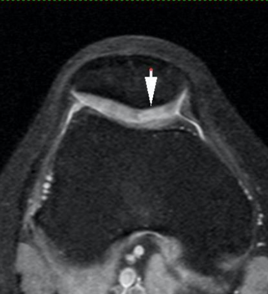
Example of MRI showing cartilage damage


This is an MRI scan image of the knee in cross-section view.
Blue outlines indicate the Kneecap bone (Patella bone) at the top, and the main knee bone (Femur bone) at the bottom.
- Red marking shows normal cartilage.
The gap between the normal cartilage is an area of damaged cartilage. In this case, the damage is Full-Thickness, so that there is now an ulcer/crater in the cartilage over the Femur.
Another MRI Example


Again, the blue outline indicates the Patella bone (top) and Femur bone (bottom), and the red marking indicates the normal cartilage.
In this case, the cartilage ulcer is over the Patella bone.
What is done in the treatment?
Patients with MRI findings such as the ones shown above will benefit greatly from Cartilage Repair Treatment.
The treatment is done using a Key-Hole procedure, where 2 to 4 small openings of 6-7mm each are made around the knee, so that thin instruments can be used to perform the treatment.
- The area of cartilage damage is first cleaned up, removing torn cartilage and leaving healthy cartilage.
The cartilage is then repaired by making small holes in the bony base and then injecting a commercially-available gel to regenerate the cartilage.
Case A - Trochlear cartilage repair


These images are of the same patient. The right image shows damaged cartilage like a crater, corresponding to the ulcer seen on the MRI.


The left image shows the defect after the damaged cartilage has been cleaned out, leaving only healthy cartilage.
The right image shows the defect filled up with the injectable gel mixed with the patient’s blood.
Case B - Patella (Kneecap) cartilage repair


These images are of the same patient. The middle image shows torn nad tethered cartilage, corresponding to the full-thickness cartilage damage seen on the MRI.
The right image shows the process of clearing out damaged cartilage using an instrument called a curette.


The left image shows the process of making small holes in the bony base, called ‘micro-fracture’.
The middle image shows the injection of the gel.
The right image shows the completed re-surfacing of the defect.
Science behind the Treatment
This treatment stimulates the repair of damaged cartilage in cases where there has been cartilage wear-and-tear or in cases of cartilage injury.
Removal of damaged cartilage is important as such damaged tissues will not heal and will continue to cause pain if left in the body.
The process of making small holes in the bony base allows the body’s own Mesenchymal Stem Cells to flow into the defect. Such cells come from marrow, or the internal soft bone.
To achieve reliable cartilage formation, the artificial gel, which is a collagen matrix, is injected into the defect and this effectively traps the Mesenchymal Stem Cells in the defect and allows the regeneration of cartilage.
Recovery of a typical case
The patient stays for one night in the hospital for rest, antibiotics, and physiotherapy treatment the next day.
Patients use the assistance of 1 crutch to walk, for about 5 to 10 days.
Patients wear a knee brace to support the knee and prevent the knee from bending too much. The brace is typically used anywhere from 1 week to 4 weeks after treatment.
Physiotherapy starts about 2 weeks after treatment and may last for 4 to 8 weeks.
Patients may commence cycling and swimming about 4-6 weeks after treatment.
Patients may resume running and sports about 12 weeks after treatment.